Macular degeneration refers to a group of progressive eye diseases that damage the macula, the central part of the retina at the back of the eye, leading to a loss of central vision. Age-Related Macular Degeneration (AMD) Dry: Also called atrophic AMD , dry AMD is the most common type, affecting around 80 percent of those with AMD. Age-related damage to a support membrane under the retina is believed to contribute to this condition.
Nearly all cases of macular degeneration begin as the dry form. Wet: Although wet AMD is less common, it is the leading cause of severe vision loss and causes most of the vision loss associated with AMD. It is also known as exudative AMD.

Non-Age-Related Macular Degeneration Stargardt macular dystrophy (STGD): Stargardt disease, also known as Stargardt macular dystrophy, is the most prevalent juvenile macular degeneration, affecting roughly one in 10,000 people. STGD is characterized by early vision impairment, often before visible changes appear in eye examinations. This may lead children to avoid chores or schoolwork and be mislabeled as slackers.
X-linked juvenile retinoschisis (XJR): X-linked juvenile retinoschisis occurs almost exclusively in males. It is the primary genetic cause of macular degeneration in boys, affecting around 1 in 5,000 to 25,000 individuals. It constitutes roughly 5 percent of all inherited retinal dystrophies starting in childhood.
XJR is typically identified when affected boys begin school, and signs of poor vision and reading difficulties emerge. Best vitelliform macular dystrophy (BVMD): Best vitelliform macular dystrophy, also known as Best disease , is a macular disease typically seen in childhood. It is characterized by a distinct deposit of orange or yellowish material, resembling egg yolk , within the macula.
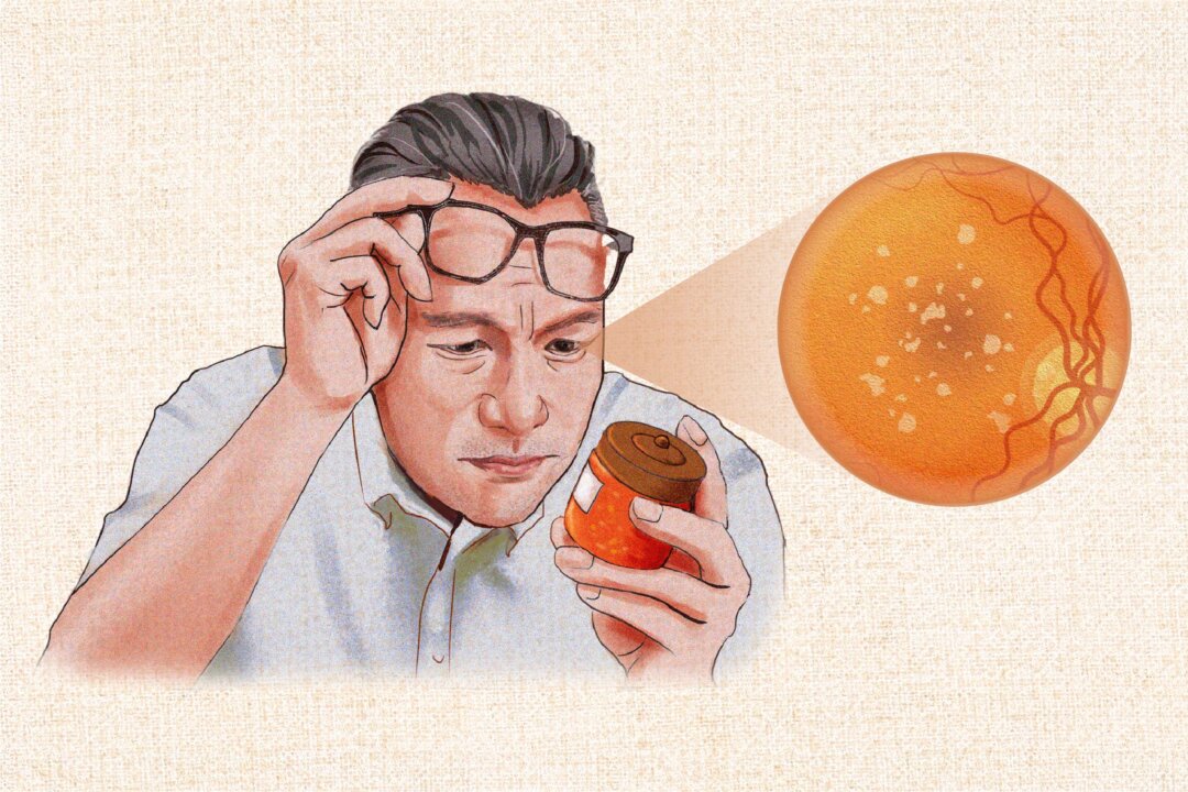
The presence of drusen is also a common early sign of AMD, especially in its dry form. Drusen are small, yellow protein deposits in the retina that can be either hard or soft. While most people over 40 have a few small, hard drusen, having many or larger ones can signal the onset of macular degeneration.
These deposits block sufficient oxygen and nutrients from reaching the retina. However, you cannot detect drusen yourself, so visiting an eye care professional with specialized tools is required. Cataracts: Symptoms, Causes, Treatments, and Natural Approaches Glaucoma: Symptoms, Causes, Treatments, and Natural Approaches Vitamin A: Deficiency Symptoms, Health Benefits, Optimal Sources, and Side Effects Blurry or fuzzy vision Challenges in identifying familiar faces Central vision decline or eventual loss Objects nearby appearing to change shape, size, or color, or move or vanish Increased light requirement for close-up work Print blurring or words disappearing during reading Diminished color brightness and fading colors Haziness or lack of clarity in vision The presence of a blurry or blind spot in the central field of vision, potentially expanding in size and darkening over time Retinal swelling Gray-green discoloration beneath the macula Leakage of fluid or deposits around the macula Detachment of retinal pigment epithelium (RPE), a layer of cells located between the retina (the layer at the back of the eye that converts light into images) and the eye’s choroid (the layer of the eye that supplies nutrients) Bleeding below the retina near the macula Objects appearing either more distant or smaller than their actual size Abrupt blind spot development Rapid vision loss Straight lines may seem wavy or bent (metamorphopsia) In dry AMD, RPE cells lose their pigment and struggle to eliminate waste from rods and cones, leading to waste buildup and eventual deterioration of the rods and cones.
As these cells die off, they are not renewed. In the wet type, abnormal blood vessels develop beneath the macula, causing fluid or blood leakage. The exact cause of this new vessel growth is unknown, although researchers speculate it may result from waste removal breakdown.
This could explain why dry AMD often precedes wet. These blood vessels impede nutrient delivery to the macula, leading to the deterioration of rods and cones. Additionally, research indicates that inflammation is a key factor in the development of wet AMD.
Age-related macular degeneration occurs when the retinal pigment epithelium becomes less efficient at supplying nutrients and removing waste. This causes the formation of drusen, protein deposits that interfere with vision. In wet AMD, abnormal blood vessel growth causes leakage, resulting in a dark spot in the center of the visual field.
(Illustration by The Epoch Times, Shutterstock) Early: Early dry AMD is characterized by more than 15 small drusen or fewer than 20 medium-sized, indistinct soft drusen (63 to 124 microns in diameter) or pigment abnormalities. It often presents no symptoms, making it difficult to diagnose. Intermediate: Intermediate dry AMD is marked by at least one large druse (over 125 microns in diameter), numerous medium-sized drusen, or noncentral geographic atrophy (atrophy not involving the fovea, a small, central pit in the retina).
Some people have no symptoms, while others may experience mild ones, such as blurriness in central vision or difficulty seeing in low light. Advanced: Advanced AMD, also called late AMD, involves central geographic atrophy affecting the fovea or wet AMD. Geographic atrophy gets worse over time and usually affects both eyes.
People who have the condition in one eye are also at risk of developing the wet form of AMD in the other eye. A common symptom of dry AMD is seeing straight lines appear wavy. See your eye doctor immediately if you notice this warning sign of late AMD.
Age: Age is the leading risk factor for macular degeneration. People aged 50 and older are at higher risk. Race: One 1999 study found that while drusen are prevalent in both black and white individuals aged 40 and above, severe forms of age-related maculopathy and late AMD are more common in older white people.
In addition, early AMD is more prevalent in people of European ancestry compared to Asians, but the prevalence of late AMD is similar in both populations. Light eye color. Sex: AMD affects both men and women equally.
However, since women generally have longer lifespans than men, they tend to be diagnosed with AMD more frequently. Diet high in saturated fat. Elevated high-density lipoprotein (HDL) cholesterol: A 2004 analysis of the Rotterdam Study , an ongoing cohort study that started in 1990, found that increased HDL cholesterol, but not total cholesterol, was associated with an increased risk of AMD.
Obesity: In a 2001 analysis of a study involving 2,584 participants, obese individuals had a 129 percent higher risk of developing late AMD and a 54 percent higher risk of experiencing pigmentary abnormalities compared to lean individuals. Smoking: Smokers have a significantly higher risk of developing macular degeneration—up to four times more than nonsmokers. In addition, the risk of developing the condition for a smoker with a common gene for AMD increases to 20 times compared to nonsmokers with the same gene.
A 2007 analysis of the European Eye (EUREYE) Study reported that current smokers have 2.6 times the risk of developing wet AMD and 4.8 times the risk of developing advanced dry AMD compared to nonsmokers.
Underlying medical conditions: Individuals with high blood pressure are almost twice as likely to develop AMD compared to those with normal blood pressure. People with cardiovascular diseases are also at increased risk. Family history: Around 15 percent to 20 percent of individuals with AMD have a first-degree family member (e.
g., parent, sibling, or child) who also has the condition. Prolonged exposure to sunlight.
Specific genes: Examples include the ABCA4, RS1, and BEST1 genes. Certain genetic variants in the complement system, part of the immune system, are linked to AMD. One well-known example is the Y402H variation in the complement factor H (CFH) gene.
Insufficient levels of antioxidants. Regular use of certain pharmaceuticals: A 2012 analysis of the EUREYE Study concluded that using aspirin often is linked to early AMD and late wet AMD, and the risk increases with more frequent use. People who regularly take antacids are also at a higher risk.
Stress: Stress may contribute to increased inflammation, and since AMD is an inflammatory disease , this connection could potentially exacerbate the condition. Sedentary lifestyle. Severe myopia: Whites, East Asians (e.
g., Chinese and Japanese), and Ashkenazi Jews have a higher prevalence of myopia than other ethnicities. First, a visual acuity test may be given.
This test uses a standard eye chart to assess the clarity of vision at different distances. Your visual field is the extent of the entire area you can see when focusing on a central point. A visual field test measures how much vision you have in each eye and tracks vision loss over time.
This test can identify blind spots and determine their size and shape, indicating the impact of eye diseases or brain disorders on your vision. There are several vision field tests. One often used to diagnose early AMD is the Amsler grid.
An Amsler grid can be used to monitor visual changes. If blank spots appear or the lines become warped-looking, it may indicate macular degeneration. (V_ctoria/Shutterstock) If an Amsler grid is unavailable, you can test for visual distortions by looking at a straight edge or right angle, such as a door frame or window, with one eye at a time.
Microperimetry: Microperimetry is a visual field test that combines perimetry (measurement of visual field function) and retinal imaging to directly map stimuli in specific regions of the retina. This relation of functional (visual field) and structural (retinal imaging) data aids in assessing vision quality in conditions such as AMD. It identifies central blind spots and links diminished macular sensitivity with the severity and progression of AMD.
Fundoscopy: Also known as fundoscopic exam or ophthalmoscopy , fundoscopy uses an ophthalmoscope or a magnifying lens and light to examine the back of the eye, including the retina and optic nerve. Pupils may be dilated with eye drops to allow the doctor a clear view. Retinal damage is often visible even before symptoms appear.
Fundus fluorescein angiography (FFA): Also known as fluorescein angiography (FA), FFA uses a special camera to capture blood flow in the retina. It is the gold standard for spotting new blood vessel growth caused by AMD. The procedure involves dilating the eyes and injecting fluorescein dye into a vein in the arm or hand.
As the dye travels through the retinal blood vessels, photographs are taken to detect abnormal vessels or damage beneath the retina. The test is noninvasive and does not involve direct contact with the eyes. Indocyanine green angiography (ICGA): Indocyanine green angiography uses indocyanine green dye to examine blood flow in the choroid, the layer of blood vessels beneath the retina.
The dye is injected into a vein in the arm or hand, and as it circulates through the eye’s blood vessels, photographs are taken to capture the blood flow. Autofluorescence imaging: Autofluorescence imaging uses the retina’s natural fluorescence to assess the health and activity of the RPE. Your eye doctor will use a blue light to illuminate the retina, causing specific cellular components to fluoresce without the need for dye injection.
The resulting black-and-white image reveals characteristic patterns that evaluate RPE activity and define areas of geographic atrophy. Atrophic areas will appear darker due to a loss of pigment cells, while their edges may look slightly brighter. Optical coherence tomography (OCT): OCT is a noninvasive imaging technology that provides detailed cross-sectional images of the retina.
It uses light rays to measure retinal thickness and the area and volume of drusen, aiding in the early detection and diagnosis of retinal diseases without the use of radiation or X-rays. OCT scans are painless and considered simpler, faster, and safer than FFA. Color fundus photography: Color fundus photography uses a specialized camera to capture color images of the eye’s interior surface.
This method helps document AMD and track its progression over time. Legal blindness: Without treatment, AMD may lead to legal blindness (vision worse than 20/200) in the affected eye. Subretinal neovascularization: Subretinal neovascularization occurs when blood vessels accumulate under the retina, potentially leading to retinal detachment (a medical emergency) or scarring.
It significantly reduces the likelihood of preserving central vision and affects about 20 percent of AMD cases. This condition frequently recurs despite treatments such as laser treatment. Charles Bonnet syndrome: Having hallucinations due to vision loss is known as Charles Bonnet syndrome.
Up to 50 percent of people with macular degeneration may experience visual hallucinations at some point. This condition is often associated with significant vision loss. Depression: People diagnosed with AMD face an increased risk of developing depression , especially if they experience visual impairment due to the condition.
Retinal atrophy: Myopia stretches the retina, resulting in thinning that can lead to areas of retinal atrophy (breakdown) and subsequent vision loss. Dry AMD AREDS or AREDS 2 formulas: People diagnosed with intermediate or advanced AMD in at least one eye may want to consider taking the dietary supplement recommended by the Age-Related Eye Disease Study (AREDS) and the AREDS 2 (a second study). The AREDS 2 formula comprises daily doses of 500 milligrams (mg) of vitamin C, 400 international units (IU) of vitamin E, 80 mg of zinc oxide, 2 mg of cupric oxide, 10 mg of lutein, 2 mg of zeaxanthin, and 1 gram of omega-3 fatty acids.
The original AREDS formula contains beta carotene, which can increase lung cancer risk in current or past smokers, so ensure you choose AREDS 2 if you have a history of smoking. Vision aids: Various types of vision aids are available, such as magnifiers, products enhancing color or contrast for better visibility, larger-font-sized items such as clocks, large-print books, reading stands, anti-glare glasses, task lights, and leisure products such as large-print playing cards and dominoes. Vision rehabilitation programs: Vision loss rehabilitation therapy assists individuals with various types of vision loss in developing or restoring essential daily living skills, improving their independence and mobility.
Medication: In 2023, two new drugs, pegcetacoplan and avacincaptad pegol, received approval from the U.S. Food and Drug Administration (FDA) for treating late-stage dry AMD.
In one 2023 study , monthly intakes of pegcetacoplan slowed the progression of geographic atrophy (late-stage dry AMD) by 21 percent. Another study showed that avacincaptad pegol could slow the condition’s progression by 14 percent. Occupational therapy.
Wet AMD Anti-VEGF medications: Vascular endothelial growth factor (VEGF) stimulates new blood vessel growth in the body. In wet AMD, excessive VEGF levels cause uncontrolled blood vessel growth in the eye. Anti-VEGF therapies inhibit VEGF activity, potentially halting or reversing vision loss.
These treatments involve injections directly into the eye and are typically the primary approach for managing wet AMD. Anti-VEGF injections usually provide only temporary relief, necessitating frequent treatments for most patients. Examples of anti-VEGF medications include aflibercept, bevacizumab, brolucizumab, faricimab, and ranibizumab.
Photodynamic therapy: In photodynamic therapy, verteporfin is injected into an arm and travels to the blood vessels in the eye. A laser is then used to activate the drug, helping reduce leakage from the blood vessels. Laser photocoagulation: Laser photocoagulation, or photocoagulation therapy, involves using a laser to seal leaky blood vessels in the eye and prevent further leakage.
This procedure creates small burns in the retina, helping seal or destroy abnormal blood vessels, prevent the growth of new blood vessels, and reduce the risk of vision loss. As a result, the retina becomes stabilized to preserve vision. Laser surgery: Laser surgery for wet AMD is a last-resort treatment to seal abnormal eye blood vessels that continue to leak despite other therapies.
While it may slow vision loss, it doesn’t restore vision and can lead to scarring and blind spots. Surgery: In rare cases where other treatments have not been effective, surgical removal of abnormal blood vessels may be considered a last resort. Levodopa: The use of levodopa (L-DOPA), a medication commonly used to treat Parkinson’s disease and a precursor to dopamine, is linked to a decreased incidence of newly detected wet AMD.
In one 2021 pilot study , levodopa reduced fluid in the retina, delayed the need for standard injections of anti-VEGF medication, and improved vision outcomes. By Month 6, patients in both study groups showed improved vision, with an average gain of 4.7 to 4.
8 letters on vision charts. Also, the need for anti-VEGF injections was reduced by 52 percent in one of the study groups. As aforementioned, stress can exacerbate inflammation, a known factor in the progression of AMD.
Thus, a negative mindset characterized by chronic stress, anxiety, or depression can increase systemic inflammation, potentially accelerating macular degeneration. On the contrary, a positive mindset can enhance a patient’s adherence to treatment regimens and influence the person to adopt healthy lifestyle choices to help slow the condition’s progression. 1.
Medicinal Herbs Saffron: According to a 2021 systematic review , in vitro studies showed that saffron , the most expensive spice in the world, could protect photoreceptors in retinal cells from light-induced damage. A 2024 study found that saffron supplements could benefit people with AMD by modestly improving eye function. This improvement was observed even in those already taking AREDS supplements.
The researchers believed that long-term supplementation might result in more robust benefits given AMD’s chronic nature. Ginkgo biloba: A 2013 review of two trials found that ginkgo biloba extract had potential benefits for vision in patients with AMD. Its mechanisms of action might include enhancing blood flow, blocking platelet-activating factors, and protecting against membrane damage from free radicals.
Turmeric: The root of the turmeric plant is commonly used as a spice in cooking, especially in South Asian cuisine. Curcumin is the active ingredient in turmeric and is known for its anti-inflammatory and antioxidant properties. In treating retinal diseases , curcumin has been shown to modulate gene transcription, reduce ganglion cell death, downregulate VEGF, and decrease vascular dysfunction.
A 2022 meta-analysis found that curcumin helped treat AMD by lowering harmful reactive oxygen species and inhibiting proteins that cause cell death and inflammation. It also boosted protective proteins. Resveratrol, quercetin, and curcumin: In a 2023 phase 2 trial involving 25 dry AMD patients who took either an oral combination of resveratrol, quercetin, and curcumin (RQC), curcumin alone, or nothing for one year, the RQC group’s average drusen volume decreased by 10 percent, while the curcumin group’s average decreased by just under 4 percent.
2. Diets Mediterranean: According to a 2022 systematic review , research consistently shows that following a Mediterranean diet pattern reduces the risk of developing AMD and the risk of advancing to more severe stages of the disease. This underscores the importance of preventive measures that promote diets rich in plant-based foods.
Ketogenic and low-protein: The keto diet, characterized by high fat and low carbohydrate intake, may improve neurodegenerative diseases, and its benefits for retinal degeneration are being researched. A 2020 animal study found that the ketogenic and low-protein diet was linked to reduced phototransduction (how light is converted into electrical signals) and lower serum albumin levels, which might act as a protective mechanism. Fatty fish: Research suggests that docosahexaenoic acid (DHA) and eicosapentaenoic acid (EPA) , omega-3 fatty acids found in retinal cell membranes and known for producing beneficial compounds, may protect the retina and potentially prevent or slow AMD.
A 2008 study involving 2,275 older participants showed that eating fatty fish at least once a week cut the risk of participants developing wet AMD roughly in half, compared to eating fatty fish less frequently. 3. Supplements Astaxanthin: Astaxanthin is a powerful antioxidant that stands out among other carotenoids, such as zeaxanthin, lutein, canthaxanthin, and beta carotene.
It is 500 times more potent than vitamin E, which is why it’s nicknamed “super vitamin E.” Studies suggest that astaxanthin has the potential to enhance eye health, demonstrating notable improvements in the management of various eye conditions, including AMD. Vitamin D: A 2017 systematic review found that vitamin D deficiencies were consistently linked to AMD and that individuals with low vitamin D levels are at a higher risk of both early and late stages of AMD.
Another 2017 study involving 2,146 participants found that people who consumed the highest amounts of vitamin D in their diet had a 40 percent lower risk of their AMD worsening than those who consumed the least. A diet rich in vitamin D may prevent or delay the progression to advanced AMD, particularly wet. Melatonin: A study published in June found that supplementing with melatonin reduced the risk of developing AMD by 58 percent.
For those who already had dry AMD, it reduced their risk of it progressing to wet by 56 percent. However, the researchers noted that their study could not prove causation and that melatonin users might generally have healthier lifestyles, which could contribute to reduced disease risk. 4.
Acupuncture Upping intake of lutein and zeaxanthin: A 2007 analysis of the AREDS involving 4,519 participants aged 60 to 80 years at enrollment found that a higher dietary intake of lutein and zeaxanthin was independently associated with a reduced risk of developing wet AMD, geographic atrophy (the advanced stage of dry AMD), and large or extensive intermediate drusen. Sources of lutein include egg yolks, leafy green vegetables, red grapes, yellow corn, and peas. Zeaxanthin can also be found in leafy greens, corn, and peas, as well as in goji berries, pistachios, and summer squash.
Eating enough fruits and vegetables: Antioxidants and other compounds in fruits and vegetables protect the body against the effects of free radicals. Avoiding smoking: After quitting smoking, the risk of developing AMD begins to decline. Twenty years later, the risk is comparable to that of someone who has never smoked.
Exercising regularly to maintain a healthy weight. Protecting eyes from overexposure to sunlight. Managing and monitoring underlying conditions, such as diabetes and hypertension.
Staying up to date with monitoring: Since early AMD doesn’t show symptoms, regular eye exams are crucial if you’re at risk of AMD. Consult your health care provider about the frequency of these exams..