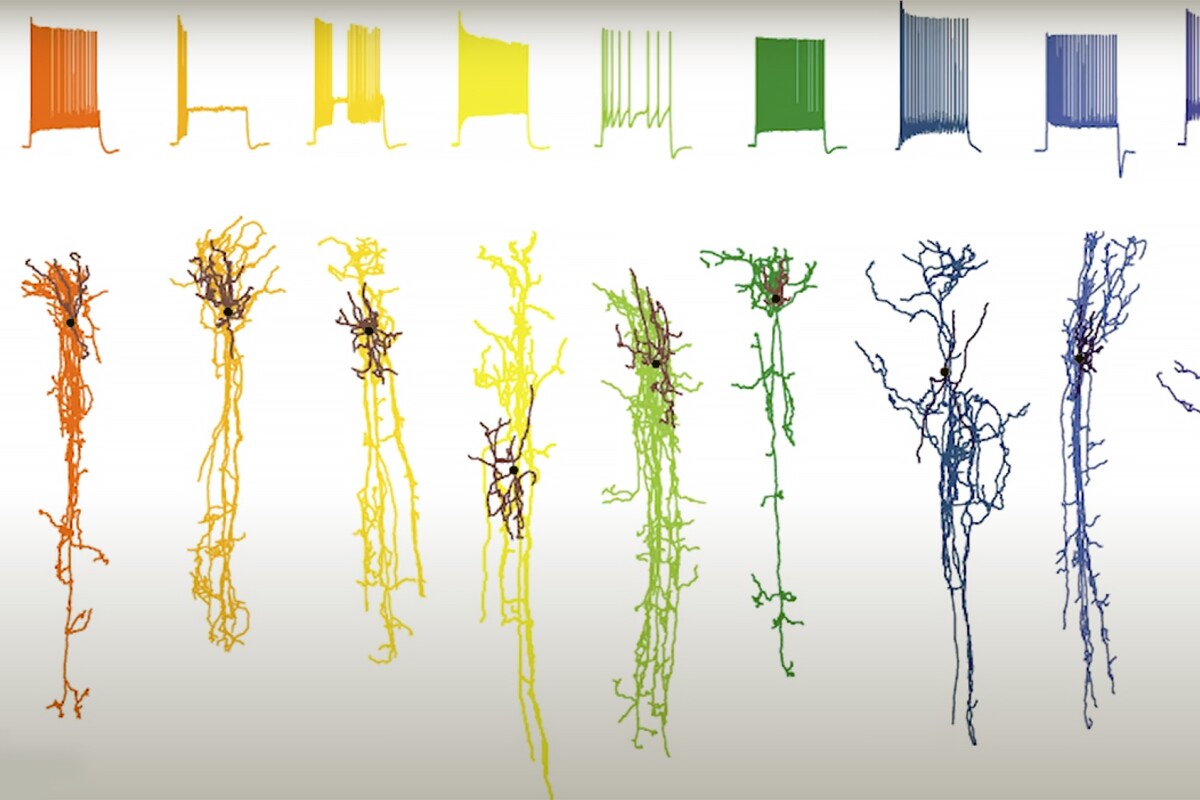
By obtaining high-resolution images of Alzheimer’s disease-affected brain cells as the condition progresses, researchers have identified the specific neurons that are damaged. The information the ‘cell atlas’ provides highlights potential new treatment targets. After identifying that accumulation of toxic and proteins in the brain are a feature of Alzheimer’s disease, researchers have, understandably, been working on treatments that reduce or eliminate them.
Less research has focused on the specific cells affected by the condition. Now, researchers from the Allen Institute for Brain Science, the University of Washington (UW) Medicine, and the Kaiser Permanente Washington Health Research Institute have created a high-resolution timeline of images that show how Alzheimer’s disease progresses at the cellular level. “The takeaway is that this atlas describes AD [Alzheimer’s disease] progression at unprecedented cellular resolution, and identifies many new cellular and molecular targets for the field to explore,” said Kyle Travaglini, PhD, a scientist at the Allen Institute and the study’s co-lead author.
The researchers zeroed in on cells from a particular region of the brain’s outer layer, or cortex, called the middle temporal gyrus (MTG), which is involved in language, memory processing, and visual perception. The MTG is also a critical ‘transition zone,’ where preclinical Alzheimer’s pathology, such as the buildup of toxic proteins, transitions to mor.


