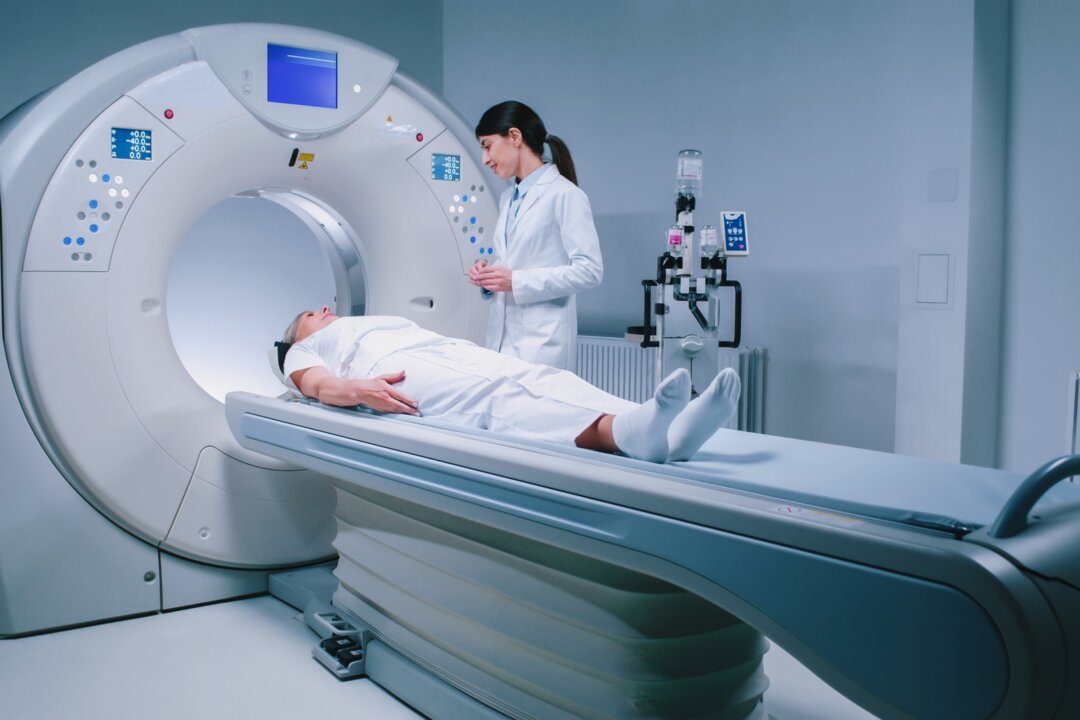
The study notes that even a single CT image from the lower back can reveal critical details about these markers, potentially predicting events like cardiovascular disease and overall mortality. The study examined how well these CT-derived markers could predict Type 2 diabetes and other related health issues. Researchers analyzed data from more than 32,000 South Korean adults who had previously undergone health screenings, including positron emission tomography (PET)/CT scans.
At the start of the study, 6 percent of participants had diabetes, and 9 percent developed it during the follow-up period. The automated CT markers were effective at predicting new diabetes cases, with accuracy scores of 0.68 for men and 0.

82 for women (where 1.0 indicates perfect accuracy). The amount of visceral fat around the abdominal organs was the best predictor of diabetes.
When combined with measures of muscle size, liver fat levels, and calcium buildup in the arteries, the predictions became even more accurate. Developed by a multi-institutional team, the AI model flagged elevated diabetes risk years before diagnosis by analyzing more than 270,000 x-rays and electronic health records, focusing on fatty tissue location. Validated on nearly 10,000 additional patients, the authors explained that this approach offers a cost-effective method for early detection.
One significant obstacle is securing reimbursement that reflects the actual value of these screenings. Without proper compensation, integrating these advanced AI tools into routine practice may be financially unsustainable, he said. Additionally, there is a risk of backlash and misunderstanding from primary care providers if the rollout is not managed correctly.
Opportunistic screening will likely identify unsuspected findings or patients at high risk for disease. Appropriate referral networks must be established to help patients and providers manage these findings. Pickhardt noted that the increased workload for radiologists could complicate this process.
“I am very concerned about the radiation risk with all x-ray-based imaging, including CT and mammography,” Semelka said. He pointed out that the radiation dose from CT scans can increase the risk of malignancy, a serious consideration for screening purposes. Semelka questions whether the benefits of CT imaging outweigh the risks and costs, primarily since methods like MRI can provide similar or better results without the cancer risk.
“How much better is this than using height, weight, BMI, and abdominal circumference to determine T2DM [Type 2 diabetes mellitus] risk?” he asked, suggesting that conventional methods may be more practical for routine screenings. Despite his concerns, Semelka acknowledges that add-on or opportunistic scans without additional radiation can be beneficial. “An add-on or opportunistic scan that does not add any additional radiation is always a good idea,” he said.
However, he criticized the use of PET-CT for general screening due to the high radiation dose. While CT imaging offers advanced insights, particularly in measuring visceral and liver fat, Semelka advocates for cautious use. He believes that imaging should not be the sole method for screening unless it is part of a comprehensive approach that includes genetic testing for those with a high predisposition to certain diseases.
“The risk of the imaging procedure should not greatly exceed the risk of the potential diseases being looked for,” he said, emphasizing the need to balance the benefits and risks of using imaging technology in medical practice..


